Researchers at Orel State University in Russia have developed a biopsy system that can differentiate between healthy and cancerous tissue in many clinical cases. The device was designed to address the difficulties doctors can have when biopsy of a liver tumor, which can make it difficult to know if the needle is in the right place for small, early-stage tumors. The system uses a combination of lifetime fluorescence measurements and diffuse reflection spectroscopy to identify a tumor.
Obtaining a tumor biopsy is an important first step in identifying its characteristics so clinicians can plan treatments accordingly. However, it can be difficult to be sure that the tissue you just removed with a hollow needle was actually from the tumor itself, especially if the tumor is small and located in the abdominal cavity.
These Russian researchers came up with a biopsy system that could help. “Optical biopsy methods like the one we developed make it possible to differentiate between healthy and tumorous tissue with great accuracy,” says Elena V. Potapova, a researcher involved in this latest study. “Although our system was developed specifically for use in abdominal surgery, our results show that similar technologies could also be useful for other medical applications.”
The technology combines two different modalities to identify tumor tissue in near real time. The first is diffuse reflectance spectroscopy, which measures how the tested tissue reflects light, and the second is called fluorescence lifetime analysis. Fluorescence is induced by shining a certain wavelength of light onto a tissue and then calculating how long it takes for the fluorescence signal to disappear.
Molecules that are involved in the cell metabolism within a tissue influence the duration of the fluorescence. Because cancer cells have significantly altered metabolism, the technique is useful for quickly determining cancer status. “Although our team and others have previously used fluorescence intensity to assess tissue, studies on other parts of the body have shown that fluorescence lifetime is less dependent on experimental conditions,” said Potapova. “Fluorescence lifetime measurements remain more consistent in the presence of blood, with uneven lighting, or when the probe-tissue contact changes due to movement.”
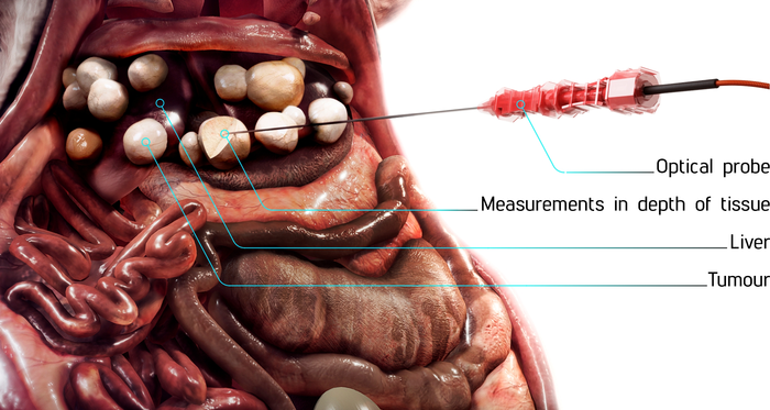
The probe is only 1 mm in diameter and the system is compatible with standard 17.5 G biopsy needles.
Studies in Biomedical Optics Express: Fluorescence lifetime needle optical biopsy differentiates hepatocellular carcinoma
Over: OPTICAL