Researchers at Harvard developed a technique that lets them recreate the helical arrangement of cardiac muscle fibers within the heart ventricles in a bioengineered construct. The breakthrough could pave the way for artificial bioengineered hearts.
The researchers used a technique called Focused Rotary Jet Spinning (FRJS) that allows them to deposit tiny polymer fibers in a helical fashion very rapidly. When seeded with cardiac cells, the resulting artificial ventricles begin to beat after a few days, and the researchers showed that this arrangement results in a greater ejection fraction and more rapid electrical signaling compared with circumferential fibers. The results highlight the importance of considering natural structures as closely as possible when creating bioengineered constructs, as hundreds of millions of years of evolution have resulted in our organs doing their job pretty efficiently.
Scientists noticed the helical arrangement of ventricular heart fibers centuries ago, and in the 1960s a researcher named Edward Sallin hypothesized that this made the heart better at pumping blood, but until now no-one could prove it. “Since 2003, our group has worked to understand the structure-function relationships of the heart and how disease pathologically compromises these relationships,” said Kit Parker, a researcher involved in the study. “In this case, we went back to address a never tested observation about the helical structure of the laminar architecture of the heart. Fortunately, Professor Sallin published a theoretical prediction more than a half century ago and we were able to build a new manufacturing platform that enabled us to test his hypothesis and address this centuries-old question.”

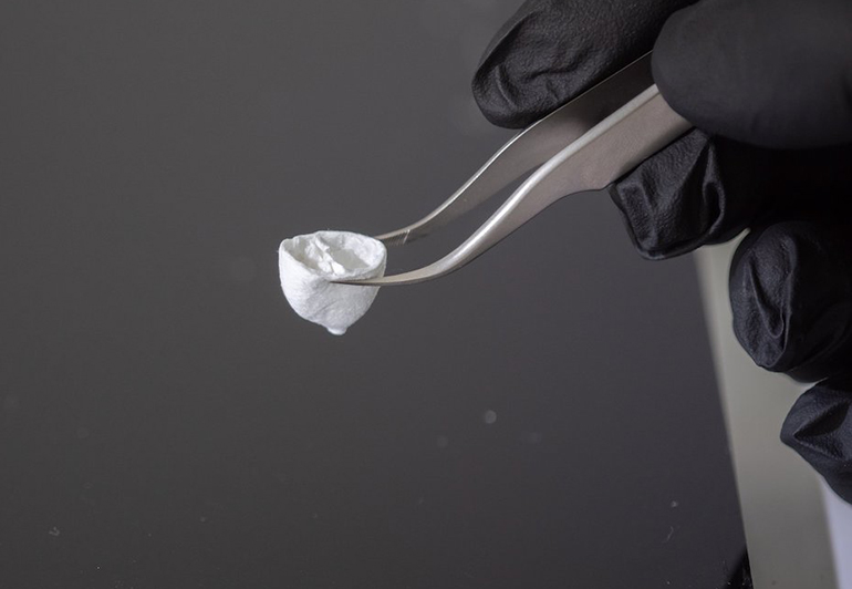
To create the artificial structures, the researchers used a new technique called Focused Rotary Jet Spinning (FRJS), which resembles the way that cotton candy is made. A liquid polymer solution is extruded from a reservoir and begins to solidify rapidly in the air. An air jet directs the polymer in the air and it is deposited onto a spinning collector. By changing the angle of the collector, the researchers could easily create a helical structure with the polymer fibers.
In this context, the technique is very rapid compared with other additive manufacturing approaches, such as 3D printing. At a single micron scale, FRJS could recreate the collagen components of a human heart within about one day, whereas it would take over 100 years to achieve the same thing with existing 3D printing methods.
Excitingly, the Harvard researchers were able to confirm the hypothesis that helical fibers contribute to the heart’s ability to pump blood efficiently. When seeded with cardiac cells, the helically-aligned ventricles began to beat, and the researchers confirmed that they outperformed ventricles with circumferentially aligned fibers in their ejection fraction, speed of electrical signaling, and ventricular deformation.
See a video about the technology below.
Studies in Science: Recreating the heart’s helical structure-function relationship with focused rotary jet spinning
Via: Harvard