Researchers at Heidelberg University in Germany used an imaging technique called soft x-ray tomography to get highly detailed 3D images of the inside of cells, including the changes that occur when the cell becomes infected with SARS-CoV-2. The approach can produce a high resolution 3D image in minutes, which is much faster than other microscopy techniques that achieve a similar level of detail.
Imaging technology is constantly improving and our understanding of the inner workings of our cells is increasing. From identifying what happens when SARS-CoV-2 enters a cell, to developing new treatments for cancer, it is important to map cellular processes to improve our understanding and pave the way for new therapies.
Current cell imaging techniques can have difficulty producing nanoscale images within a period of time, slowing research. “Scanning electron microscopes are preferred for cell imaging because they deliver extremely sharp images on the nanoscale,” says Venera Weinhardt, a researcher involved in the study. “But this technology takes a week to scan a single cell. It also generates a tremendous amount of data that is daunting to analyze and interpret. With soft X-ray tomography, we get usable results within five to ten minutes. ”
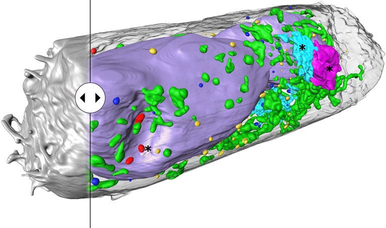
So far, the Heidelberg team has used the technology to examine lung and kidney cells infected with SARS-CoV-2 and have been able to identify changes in certain cell organelles as a reaction to an infection as well as clusters of virus particles on the cell surface. In fact, they were able to identify organelles that the virus had hijacked in order to reproduce. Fortunately, the technology works in “fixed” cells, that is, cells that have been immersed in a preservation solution that deactivates the virus particles and makes sample processing safer.
The research team was able to obtain unprecedented 3D images because they changed the design of the sample holder from a flat grid to a cylinder. The flat grids were not optimal as they often caused blurry artifacts and did not allow samples to be scanned from every angle.
“To get around this problem, we switched to cylindrical, thin-walled glass capillaries to hold the samples. During microscopy, the samples can be rotated a full 360 degrees and scanned from all angles, ”says Weinhardt.
Study in Cell Reports Methods: Use of soft X-ray tomography for rapid quantitative whole-cell imaging of SARS-CoV-2 infected cells
Over: University of Heidelberg