A team at Florida Atlantic University has developed a 3D-printed replica of a portion of the human spine, based on patient CT data. The patient-specific construct can be attached and articulated by a robotic platform, and a soft magnetic sensor allows the researchers to gauge intervertebral loads as the spine assumes different postures. The model also lets the researchers insert an artificial disc implant and measure what effect that has on the movements and intervertebral loads of the spinal replica. The technology could greatly aid spinal surgeons in planning surgical interventions in advance.
At the moment, in taking the decision to recommend a cervical disc implant, surgeons often have very limited information to guide their choice. This information typically includes imaging data, which may not provide the full picture, and at present surgeons have no real way to accurately gauge the biomechanical effects of the implant they are considering. This can have unfortunate consequences, in the form of implant failure and repeat surgeries to correct an issue.
The new technology lets a surgeon create a patient-specific 3D-printed replica based on CT data, and then test how the spinal structure moves and articulates with an artificial disc implant in place. A robotic arm works to articulate the replica, whereas a soft magnetic sensor provides data on intervertebral loads, in conjunction with machine learning algorithms.

“A flexible magnetic sensor array is a new method to realize soft and stretchable magnets by mixing silicone with magnetic powder,” said Erik Engeberg, one of the lead developers of the new technology. “These sensors are low-cost, highly sensitive, and easily integrated into robotic systems as the soft medium can be manipulated in many shapes and sizes.”
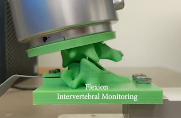
So far, the robotic replica has shown potential in assessing five different spinal postures (mid-extension, extension, center, mid-flexion and flexion), generating data that could highlight potential problems with a given implant.
“This new approach has a powerful potential to enable surgeons to preview and compare the effects of different surgical interventions in a patient-specific manner using robotically actuated spine twins,” said Frank Vrionis, another researcher involved in the study. “Moreover, the novel system could help in determining whether a constrained, semi-constrained, or unconstrained device could be the best fit or even a fusion device. Following surgery, the spine replica could also assist us in estimating whether there is sufficient motion at the operated level and possibly helping us to determine if we need to change the rehabilitation program to prevent calcification and subsequent loss of intended motion.”
See a video about the system below:
Study in sensors: Robotic Replica of a Human Spine Uses Soft Magnetic Sensor Array to Forecast Intervertebral Loads and Posture after Surgery